
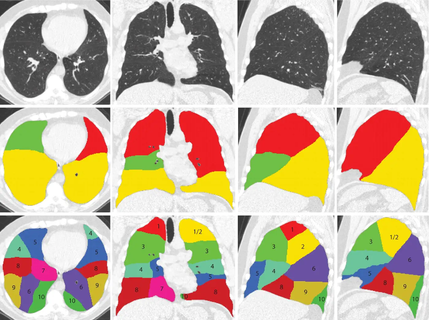
The lung presents a difficulty few organs share: the boundary that must be drawn often does not exist. A study on a corpus of chest CT reports that 147 studies were found to have at least one incomplete fissure, most frequently the right minor fissure, while accessory fissures were detected in 19 scans.An incomplete fissure means the two lobes it is supposed to separate are anatomically continuous across part of their surface. There is then nothing to see on the image, and the annotator must nevertheless produce a limit. That limit is an inference based on the probable trajectory of the fissure, not an observation.This article covers what is specific to lung segmentation: the hierarchical ontology, the fissure problem, parenchymal delineation, nodules and diffuse disease. It extends the complete guide to medical image segmentation.
A hierarchical ontology, specific to lung medical image segmentation
The lung’s structuring particularity is that its objects nest, which clearly distinguishes this domain from the organs covered elsewhere.Three levels coexist. The parenchyma, meaning the lung as a whole, separates the organ from the mediastinum and the chest wall. The lobes subdivide it, five in total, three on the right and two on the left. Lesions overlay the first two levels, a nodule necessarily belonging to a lobe and diffuse disease potentially crossing several.That hierarchy has two practical consequences. The first is a consistency constraint: every lobe voxel must belong to the parenchyma, and every lesion voxel to a lobe. That constraint is programmatically verifiable and is the field’s most effective control.The second is that the three levels do not belong to the same decision register. Delineating the parenchyma is a perceptual task based on contrast. Delineating the lobes is anatomical inference. Qualifying a lesion is a clinical decision. A protocol treating them uniformly fails on at least one of the three.Lobes and the fissure problem
This is the lung’s own difficulty in medical image segmentation, and it differs in nature from anything encountered elsewhere.Incompleteness
The figure cited in the introduction is not a recruitment anomaly: fissural incompleteness is a frequent anatomical variant in the general population. The right minor fissure is particularly exposed.The annotation consequence is direct. Where the fissure is complete, the limit is visible and can be drawn. Where it is incomplete, it must be reconstructed by continuity, extending the visible trajectory or relying on indirect landmarks, notably vascular and bronchial distribution, each lobe having its own branching.That reconstruction must be governed by a written rule. Two annotators freely extending a fissure will produce divergent limits across substantial surfaces, and the gap will show directly in lobar volumes, which are generally the quantity sought.Indirect landmarks
One method deserves teaching explicitly rather than leaving to discovery: using internal structures as landmarks. Each lobe has its own bronchial and vascular branching, and a vessel crossing a supposed boundary signals a drawing error. That cross-check is the best way to control a reconstructed limit, and it takes hours to learn. An annotator unaware of it reconstructs incomplete fissures by pure geometric interpolation, which is markedly less reliable.Accessory fissures
The opposite problem exists: an accessory fissure creates a visible limit corresponding to no subdivision in the standard nomenclature. An unwarned annotator will follow it spontaneously, producing a fractured lobe or a displaced limit.The protocol must therefore mention that variant explicitly and state that a visible limit is not necessarily a lobar limit, which is counterintuitive and must be illustrated with examples.The methodological consequence
Lobar segmentation is a case where performance depends less on contrast than on anatomical knowledge. An annotator trained to recognise normal fissural trajectories and their variants will produce better results than a more dexterous but untrained annotator, which is not the case on most contouring tasks.The parenchymal limit
Delineating the lung as a whole looks trivial given the contrast between air and soft tissue. It nonetheless holds three difficulties.The first is proximal extent. A reference protocol fixes it precisely: the mask follows the lung boundary against the mediastinum and soft tissue chest wall, and covers the lung parenchyma from the division of the primary to secondary bronchus onwards. Without that rule, whether proximal airways are included varies between annotators.The second is the diseased lung. A consolidated area has soft tissue density and therefore disappears from any air-based thresholding. The same protocol settles it: the mask includes any areas of consolidation, collapse and other abnormalities. It is the field’s most important rule, because it inverts intuition: a diseased lung is still lung, and excluding it produces a distorted denominator for any later quantification.The third is the neighbourhood. Pleural effusion, mediastinal structures and the diaphragm form sometimes blurred limits, and the protocol must state which are excluded. The same standard explicitly excludes pleural effusion from its masks, which is a decision rather than a self-evident choice.Nodules and classification variability
The nodule is the field’s most studied object, and also the one where expert variability has the most direct consequences.A study across eight radiologists and one hundred and sixty nodules reports results worth knowing: inter and intra-observer agreement is moderate, with mean kappa of 0.51 and 0.57 respectively, a discordant classification being found in 36.4 per cent of reading pairs, related to the presence or size of a solid component in 88.7 per cent of cases; two thirds of these discrepant readings would have potentially resulted in different nodule management.That result carries more force than usual variability measurements, because it concerns not an abstract score but the clinical decision that follows. More than one disagreement in three, and among those two in three changing the course of action.Two consequences for an annotation project. The first is that nodule classification does not delegate to a generalist annotator, the difficulty lying precisely in judging the solid component. The second is that a nodule classification corpus built on a single reading carries an uncertainty nothing signals, and double reading is justified there far more than on contouring tasks.Delineating the nodule
Nodule contouring itself poses three distinct difficulties. Nodules abutting the chest wall or a vessel have no visible boundary on the contact face, and the limit must be conventional. Ground-glass nodules have a gradual boundary by nature, which brings them close to infiltrative lesions. And part-solid nodules require distinguishing two components whose internal limit is precisely the point of disagreement documented above.Those three cases must appear in the protocol with examples, and they justify stratified metrics: a global score on a nodule corpus mixes unrelated difficulties.Diffuse disease
A second family of lesions behaves differently and calls for different handling.Ground glass, consolidation, reticulation, honeycombing, emphysema: these are not objects but states of the parenchyma. They have neither shape nor boundary of their own, they extend irregularly and they frequently mix.Two medical image segmentation strategies exist and the choice is structuring. Per-class segmentation distinguishes each pattern, which is clinically rich and expensive, the boundary between patterns being itself ambiguous. Binary segmentation groups everything under a single abnormality class, which is markedly more reproducible.The reference protocol already cited explicitly adopts the second approach: for the ground truth of the abnormality, a single label mask included all areas of ground glass opacity and consolidation within both lung fields. That choice deserves serious consideration, since it trades semantic richness for a reproducibility fine distinction cannot achieve.What a reference protocol documents
The same document describes its medical image segmentation organisation with rare precision, and it is worth examining because it matches the commonest production model.It states that the task was performed by a team of twenty-five expert medical annotation engineers with a median of 2.25 years of experience, under the supervision of three board-certified radiologists, every dataset being initially annotated by the annotators and later reviewed and corrected by a radiologist.That description validates a model: specialist annotators produce, clinicians supervise and correct. It also indicates that the experience required is measured in years on this kind of task, which argues for team stability.Two further elements of the same document are directly usable. Lobar masks were initialised by a pre-trained model, the annotator working in correction. And the average time for annotation and review of lung and lobe masks is given as two hours, a useful order of magnitude for costing and rarely available publicly.Airways and vessels
A fourth level appears on some medical image segmentation projects and follows its own rules: the branching structures within the lung.The bronchial and vascular trees pose a difficulty other thoracic structures do not, that of thin ramified objects. Three consequences follow, common to all structures of this type.The first is that volumetric overlap is a poor guide. A bronchial segmentation can post a respectable score while capturing only the proximal generations, which hold most of the volume, while missing all distal branching entirely. The relevant metric is tree length recovered or number of generations reached, not overlap.The second is that annotation depth must be fixed explicitly. To which bronchial generation does one descend, and by which stopping criterion: measurable diameter, lumen visibility, or number of divisions from the origin. Without that rule the work has no defined end.The third is that connectivity becomes a first-order requirement. A tree fragmented into isolated components is unusable for any path analysis, while it may show acceptable overlap. That check belongs in the automatic control.Windowing, an underestimated parameter of thoracic medical image segmentation
One technical particularity of the chest deserves treatment, since it determines what the annotator sees.The range of densities present in a chest is such that no single display setting shows everything correctly. A setting suited to the parenchyma flattens soft tissue, a setting suited to the mediastinum hides lung detail.The protocol must therefore specify the display setting to use for each structure. The document cited fixes explicit values for its task, and states that the abnormality mask was adjusted to a parenchymal window for best visualisation of the opacities.This is a point projects frequently underestimate. Two annotators working under different settings do not see the same image, and their disagreements are wrongly attributed to the protocol or to their competence. Fixing windowing per task is a zero-cost measure that removes an entire source of variability.Quality control of a lung medical image segmentation
The hierarchical structure supplies automatic checks few domains permit.Four verifications are easy to program. Inclusion, every lobar voxel having to belong to the parenchyma and every lesion voxel to a lobe. Partition, the sum of the five lobes having to reconstitute the parenchyma with no gap or overlap. Volumetric plausibility, the ratio between lobes following known anatomical proportions whose large deviation signals a fissural limit error. And laterality, a right lobe not being able to sit mostly on the left.To these is added a check specific to the domain: reviewing lobar limits in the coronal plane. A fissure drawn slice by slice in axial can show irregularities invisible in the working view and obvious in coronal, where the fissural surface appears as one piece.On lesions, control must be stratified by type. A global score mixes well-defined solid nodules with diffuse disease of gradual boundary, whose difficulties are unrelated.Annotator profile and role allocation
The hierarchy of lung objects translates directly into a hierarchy of competences, and that is what makes these projects economically viable.Parenchymal delineation is a simple perceptual gesture, largely assisted by automatic pre-annotation, and delegates without difficulty. Lobar delineation requires precise anatomical knowledge, notably of normal fissural trajectories and their variants, but that knowledge is perfectly teachable and does not involve clinical judgement. Lesion delineation requires distinguishing an abnormality from an artefact or a normal structure, which transmits through a case library. Lesion qualification, by contrast, belongs to the radiologist, and the variability figures cited above show it already poses them difficulty.That gradation permits an efficient organisation. Three of the four levels delegate to trained annotators, the clinician intervening on protocol definition, arbitration of escalated cases and qualification. It is exactly the arrangement described by the reference protocol cited above, with its twenty-five specialist annotators and three supervising radiologists.The costing consequence is significant: the radiologist time required grows not with corpus volume but with the number of lesion qualifications and contested cases, an order of magnitude lower.What the corpus must cover
The composition of a lung medical image segmentation corpus follows a particular requirement, flowing directly from the anatomical variability described above.A corpus of subjects with complete fissures produces a model that fails precisely where the lobar limit is difficult, which is where help would be most useful. The authors of a reference dataset anticipated this, selecting their studies so that both challenging and clearly visible fissures were well represented.Three coverage dimensions deserve explicit verification. Fissural state, with a sufficient proportion of incomplete fissures and some accessory fissure cases. Pathological state, an emphysematous or fibrotic lung profoundly altering parenchymal appearance and fissure visibility. And acquisition parameters, slice thickness directly conditioning the detectability of thin structures and small nodules.That verification happens when studies are selected rather than afterwards, since an already annotated corpus cannot be rebalanced without additional work.Cost and what governs it
The cost profile of a lung medical image segmentation project depends almost entirely on the hierarchical level requested.The parenchyma alone is inexpensive, contrast doing most of the work and automatic pre-annotation being highly effective. Lobes cost markedly more, reconstructing incomplete fissures not automating reliably. Lesions cost more still, and their cost varies by a large factor depending on whether they are well-defined nodules or diffuse disease.Two variables modulate that ordering. The proportion of pathological cases, a corpus from a respiratory department carrying an abnormality density that multiplies time per study. And the granularity of the lesion ontology, distinguishing between diffuse patterns costing several times what grouping under a single class does.The two-hour per study benchmark for lung and lobes, cited above, is a useful estimation base, to be adjusted for the proportion of incomplete fissures and any addition of the lesion level.Screening corpora and their particularity
A substantial share of lung projects draws on screening data rather than routine clinical imaging, and that origin carries consequences worth anticipating.Screening studies are acquired at low dose, which raises noise and degrades the visibility of exactly the structures that matter: thin fissures, small nodules, subtle ground glass. A protocol written on diagnostic-quality images will demand distinctions the screening data does not support.The prevalence profile also differs. Screening populations are largely asymptomatic, so most studies contain nothing, and the findings that do appear cluster at small sizes near the detection limit. A corpus drawn from screening therefore has an abundance of normal parenchyma and a scarcity of unambiguous lesions, which is the opposite of a clinical corpus and calls for a different sampling strategy.Two practical measures follow. Fix the acquisition dose range the protocol applies to, and exclude or flag studies outside it rather than annotating them under rules built for better images. And size the lesion-level effort from expected prevalence rather than from study count, since a thousand screening studies may yield fewer annotatable lesions than a hundred clinical ones.The most common mistakes
These failures recur often enough across lung projects that naming them is usually enough to avoid them.- Providing no rule for reconstructing incomplete fissures.
- Following an accessory fissure as though it were a lobar limit.
- Excluding consolidated areas from the parenchymal mask.
- Leaving the proximal extent of the mask towards the airways implicit.
- Not stating how pleural effusion is handled.
- Delegating nodule classification to non-specialist annotators.
- Building a nodule classification corpus on a single reading.
- Not fixing the display windowing per task.
- Reporting a global lesion score mixing nodules and diffuse disease.
- Not exploiting the available hierarchical consistency checks.