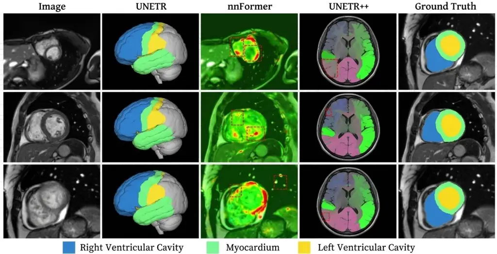

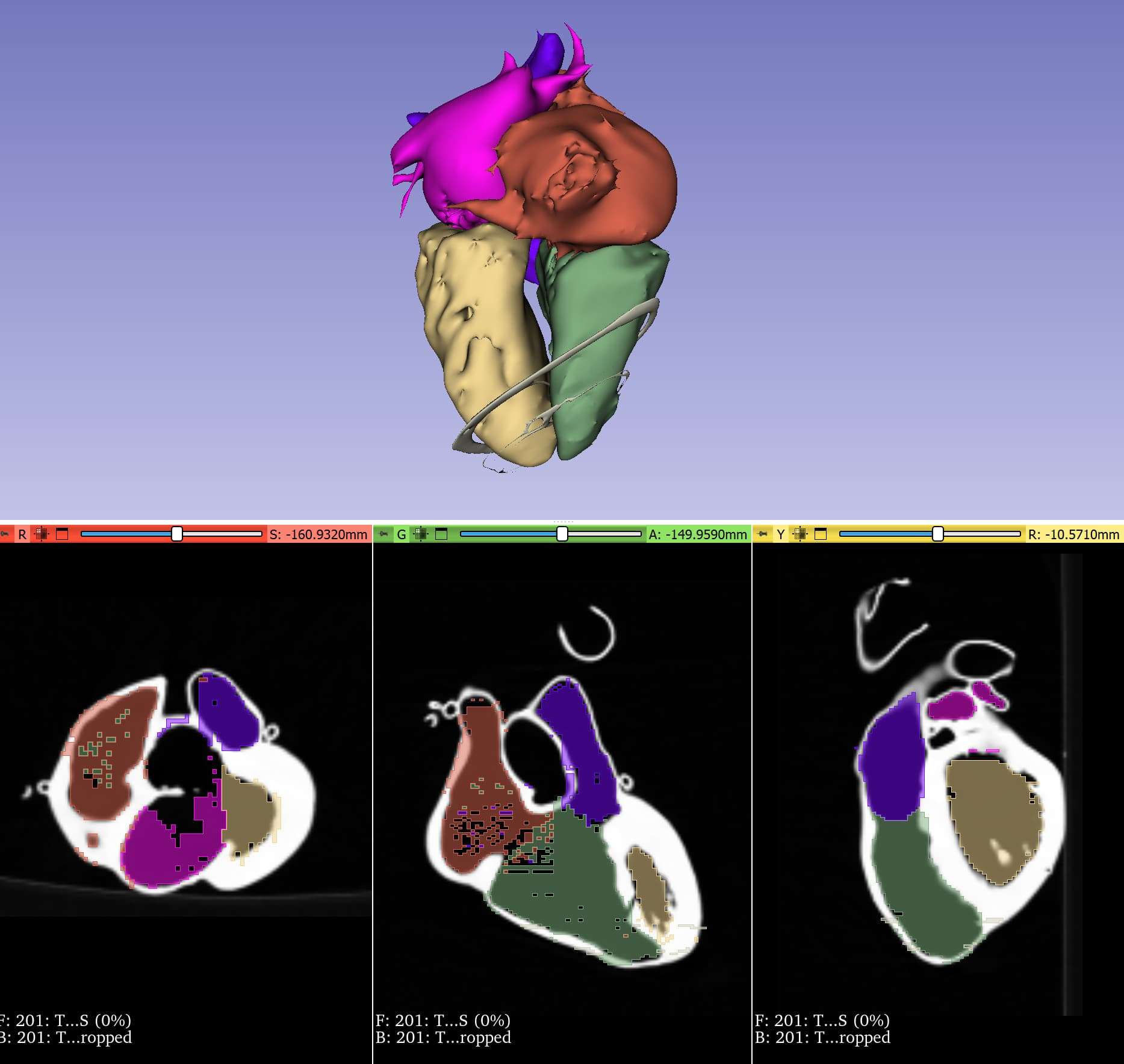
One anatomical detail captures the nature of the cardiac problem. The papillary muscles and trabeculations sitting inside the ventricular cavity have the same intensity profile as the myocardium. They resemble it because they are made of it: muscle bundles projecting into the blood pool.Including them in the cavity or in the muscle mass is therefore not a question of perception but of convention. A reference challenge protocol settles it explicitly: papillary muscles are included in the cavity and left ventricular endocardial contours follow the limit defined by the aortic valve at the base. Another convention would be equally defensible, would produce different volumes, and therefore a different ejection fraction for the same patient.This article covers that particularity: in cardiac imaging the boundary is largely conventional, and the protocol determines the clinical result. It covers structures, phase selection, extent and control. It extends the complete guide to medical image segmentation.
Three medical image segmentation questions, not one
On a fixed organ, segmenting means answering a single question: where the boundary runs. In cardiac imaging three questions arise simultaneously and each has its conventions.The first concerns structures: what exactly is delineated, and where each compartment stops relative to its neighbours. The second is temporal: the heart beats, and the same slice exists at dozens of instants in the cycle, so which ones to annotate must be chosen. The third concerns extent: the slice stack covers more than the ventricle, and where the structure begins and ends along the axis must be defined.These three are independent and each can be mis-set without the others being. A cardiac segmentation protocol that does not treat them separately produces variability no spatial metric explains.Structures and their conventions
Four structures form the backbone of most cardiac medical image segmentation projects, and their mutual definition matters as much as their individual one.The left ventricular cavity
Bounded by the endocardium, it serves volume and ejection fraction computation. Its main difficulty is the papillary convention noted in the introduction. Including them in the cavity, as the dominant convention does, simplifies drawing and slightly overestimates volume; excluding them gives a more anatomically faithful volume but a far longer and markedly less reproducible contour.The important point is that this convention must be constant across the corpus and documented, otherwise volumes are comparable neither between patients nor between studies of the same patient.The myocardium
It is defined by subtraction between epicardium and endocardium, making it a structure whose quality depends on two contours rather than one. An error on either propagates directly into the computed myocardial mass.Its own difficulty lies in low contrast with surrounding structures on several faces, and in its thinness at contraction, where measured thickness becomes sensitive to resolution.The right ventricle
This is the field’s hardest structure, for geometric reasons. Its shape is irregular and variable, its wall thin, and its boundary with the left ventricle raises a specific question. A dedicated challenge protocol settles it explicitly: on the septum specifically, the convention is not to include the interventricular septum in the right ventricular mass.That rule is arbitrary in the sense that the septum physiologically belongs to both ventricles. Settling it in writing is indispensable, since two annotators left to themselves will produce right ventricular masses differing by tens of per cent.Atria and vessels
Less frequently requested, they raise questions of limits with the afferent veins and with the valves. Where they appear in the protocol, the rule must state where the atrium stops and the veins begin, a limit with no visible existence.Choosing the phases
The temporal dimension is what most sharply distinguishes cardiac work from the rest of medical image segmentation, and it calls for its own conventions.A cine acquisition typically contains several dozen frames per cycle, and annotating them all would be as costly as it is pointless. Practice is to annotate only two instants, maximum filling and maximum contraction, from which the extreme volumes and the ejection fraction follow.They still have to be defined. A reference protocol does so with two distinct rules: end-diastole is defined as the first temporal image of each stack, that is the first cine phase of the triggered acquisition, whereas end-systole is defined on a mid short axis slice as the image with the smallest ventricular cavity area.The asymmetry between those two definitions is instructive. The first is technical and automatic, the second requires observation and judgement. It is the second that introduces variability, two annotators possibly selecting adjacent frames, and that variability feeds directly into the end-systolic volume and therefore the ejection fraction.One practical consequence follows: the phase must be chosen once on a reference slice and applied to the whole stack, rather than determined slice by slice. Letting each slice find its own end-systole produces an incoherent volume, aggregating different instants of the cycle.Extent along the axis
The third medical image segmentation question is the most neglected and yet weighs heavily on volumes.A short axis stack crosses the heart from base to apex, and which slices belong to the structure must be decided. The same protocol supplies a rule anchored in anatomy: the basal slice is inferred from the position of the tricuspid annulus as defined on the four-chamber view.That rule illustrates an important characteristic of the field: defining a limit on one view relies on another view. The annotator therefore cannot work on the short axis stack alone, and the tool must support cross-referencing.The basal slice is the main driver of volumetric variability. Near the base the cavity is wide, and including or excluding one slice changes the volume by several per cent. The difficulty worsens at end-systole, the valve plane moving apically during contraction, so the basal slice is not the same at both phases. A protocol that does not say so leaves each annotator to decide, and the disagreement will show in the ejection fraction.Why the convention determines the clinical result
This point deserves stating explicitly, because it changes how a project must be scoped.Ejection fraction is computed as the relative volume change between the two phases. It is therefore sensitive to any convention affecting either volume, and particularly to those affecting them unequally.Three conventions have that effect. Including papillary muscles adds a roughly constant volume at both phases, which raises the end-systolic volume proportionally more and lowers the computed fraction. The basal slice choice, if it differs between phases, acts directly on the difference. And the end-systole definition, if taken one frame too early or late, shifts the minimum volume.The consequence for a project is that reproducibility outweighs absolute accuracy. A corpus segmented under a constant convention, even a debatable one, yields ejection fractions comparable between patients and clinically usable. A corpus segmented with anatomical accuracy but no consistency yields measurements whose variation reflects the annotator as much as the patient.Modalities and their constraints
Three modalities occur in cardiac medical image segmentation and they do not call for the same protocol.Cine magnetic resonance imaging is the reference for quantification. It offers good contrast between blood and myocardium and complete volumetric coverage. Its difficulties are respiration, acquisitions being breath-held with possible offsets between slices, and rhythm, arrhythmia degrading the gating.Echocardiography is the most widespread modality in practice and the hardest to annotate. Contrast is low, noise substantial and part of the wall frequently obscured by shadows. The field’s corpora accordingly define phases by observed valve landmarks rather than by a temporal index, which makes selection dependent on image quality.Computed tomography offers excellent spatial resolution and limited temporal coverage. It serves coronary anatomy and valve structures more than functional quantification, which shifts the requirement towards geometric precision.A protocol does not transfer between modalities. Structural conventions can be retained, but phase selection rules and image quality criteria must be rebuilt.Annotating beyond the two phases
One development in cardiac medical image segmentation deserves flagging, since it changes the economics of some projects: growing interest in annotating the full cycle rather than its two extremes.Two phases suffice for volumes and ejection fraction. They do not suffice for anything dynamic: filling velocity, regional deformation, asynchrony between wall segments. Those measurements require segmentation across the whole cycle, multiplying annotation volume substantially.Two strategies make that cost bearable. Temporal propagation, where the annotator handles two phases and lets automatic tracking produce the intermediates, exploits the fact that cardiac motion is regular and periodic. And sparse annotation across the cycle, where only a few distributed phases are treated, suffices for many dynamic measurements.Both introduce a temporal consistency requirement absent from two-phase annotation: a structure’s area must evolve continuously through the cycle, and a discontinuity signals an error. That check, simple to program, is the control specific to this regime.The imbalance between phases
One empirical observation is worth knowing before setting a performance target.A study on right ventricular segmentation notes that both manual and semi-automatic segmentations performed better at the end-diastolic phase than at the end-systolic phase.The explanation is geometric. At contraction the cavity is small, the wall thick and internal structures prominent, which increases the relative share of the boundary and makes drawing more sensitive. Overlap metrics mechanically penalise small structures, so the same care produces a lower score.Two consequences follow. Metrics must be reported per phase rather than aggregated, otherwise different difficulties are being compared. And a uniform performance commitment across both phases is harder to hold at end-systole, which argues for a differentiated commitment.Quality control of a cardiac medical image segmentation
Physiology supplies powerful automatic checks, an advantage few domains offer.Four verifications are easy to program. Consistency between phases, end-diastolic volume having to exceed end-systolic volume, an inversion signalling a phase selection error. Ejection fraction plausibility, a value outside physiological bounds indicating a convention or extent error rather than pathology. Continuity of the per-slice area curve, an abrupt change between neighbouring slices signalling an inclusion or omission. And consistency between compartments, the volumes ejected by both ventricles having to be comparable absent particular pathology.That last check is particularly effective because it crosses two independent segmentations. A large discrepancy between right and left ejected volumes, in a patient without shunt or valve regurgitation, almost always indicates a segmentation error rather than a clinical reality.These checks do not replace review but they direct sampling, which is the main efficiency lever on a large corpus.Corpus composition
One feature of cardiac corpora deserves flagging because it bears directly on representativeness.The field’s reference datasets are built by deliberately balancing clinical categories, one reference corpus distributing its patients across healthy subjects, dilated cardiomyopathy, hypertrophic cardiomyopathy, heart failure with infarction and right ventricular abnormality.That construction is not incidental. These conditions profoundly alter geometry: dilated cardiomyopathy produces a wide cavity with a thin wall, the hypertrophic form produces the opposite, and the segmentation difficulties differ radically between them. A corpus of healthy subjects alone produces a model that fails precisely on the patients for whom the measurement matters.The practical consequence is that stratifying a cardiac corpus is done by clinical category rather than by study count, and that metrics must be reported per category. An average score on a balanced corpus masks weak performance on the most deformed category.The annotator profile
The competence required in cardiac medical image segmentation combines two registers few domains bring together.The first is perceptual: distinguishing myocardium from blood on sometimes weak contrast, recognising a wall obscured by an artefact, judging a blurred boundary. That register is acquired through practice as elsewhere.The second is procedural and it weighs more heavily here. A cardiac annotator must apply without fail a series of conventions that follow from no observation: papillary assignment, septal handling, phase selection, limit slice determination. An annotator excellent perceptually but loose on conventions produces an unusable corpus, whereas the reverse remains serviceable.Two practical consequences. Training must focus first on the conventions, with a case library illustrating each rule and its edge cases, rather than on the gesture. And qualification before production benefits from covering a complete study, phases and limit slices included, rather than a few isolated contours, since it is the whole chain of decisions that must be assessed.Cost and what governs it
The cost profile of a cardiac project is distinguished by an unusual multiplier.Each study contains several slices, two phases and several structures, quickly producing dozens of contours per patient. That multiplication is the dominant factor, more than the difficulty of any individual contour.Three variables modulate it. The number of structures requested, the right ventricle costing appreciably more than the left at equal quality. The number of phases, annotation limited to the two extremes costing a fraction of full-cycle annotation. And the modality, echocardiography demanding markedly more time per contour than magnetic resonance imaging because of noise and obscured regions.The useful estimation method times per structure and per phase on a pilot batch spanning the clinical categories, then weights by the expected composition. A costing built on healthy subjects systematically underestimates clinical corpora.Aligning the protocol with departmental practice
One final point conditions acceptance of the corpus by the clinicians who will validate it.Cardiac conventions are not universal. Professional recommendations leave latitude, and each department develops stable habits on papillary assignment, on the basal slice and on phase selection. A corpus built under a convention different from the department’s produces measurements that do not match its reference values, which is enough for it to be rejected whatever its intrinsic quality.The useful approach is therefore to ask about existing practice before writing the protocol, rather than adopting a reference challenge’s convention by default. Where the two diverge, the choice must be conscious and documented, and it benefits from following the department wherever the corpus feeds a local clinical use. Following it is also a way to secure the referent’s availability, since they recognise their own practice in the protocol rather than having to adapt to an imported norm.Delivering measurements alongside masks
One delivery decision specific to cardiac medical image segmentation is worth settling early: whether the deliverable is the masks alone or the masks plus the derived measurements.Delivering masks alone looks cleaner and pushes computation downstream. It also pushes the conventions downstream, since whoever computes the volumes must decide how to handle partial slices at the base and apex, and those decisions change the result as much as the contours do.Delivering computed volumes and ejection fractions alongside the masks costs little and has two benefits. It fixes the computation conventions in one place rather than leaving them to be reinvented. And it makes the physiological consistency checks part of production rather than something the client discovers later, since a corpus cannot be delivered with an implausible ejection fraction if the figure is computed as work proceeds.Where the deliverable includes measurements, one point needs stating explicitly: whether the volume computation treats slices as prisms of thickness equal to the slice spacing, or interpolates between contours. The two give different answers on the same masks, and disagreement over which was used accounts for a fair share of the discrepancies that surface at acceptance.The most common mistakes
These failures recur often enough across cardiac projects that naming them is usually enough to avoid them.- Not documenting the convention on papillary muscles and trabeculations.
- Leaving the handling of the interventricular septum implicit.
- Determining the contraction phase slice by slice rather than once per study.
- Not stating that the basal slice differs between the two phases.
- Defining the basal slice without relying on a complementary view.
- Reporting metrics aggregated across both phases.
- Favouring anatomical accuracy over consistency of conventions.
- Transposing a protocol between modalities without revising phase rules.
- Building a corpus with no stratification by clinical category.
- Not exploiting the available physiological consistency checks.