
A recent publication on a new medical annotation tool states in one sentence the problem most projects encounter. Of the field’s established environments, its authors note that these environments are powerful, but repetitive annotation may still need additional tooling to standardise navigation, prompt-based propagation, and quantitative export across many cases.The same document notes elsewhere that it still requires substantial manual work and careful data handling to produce consistent 3D labels at cohort scale.Those two observations pinpoint exactly the gap between two uses routinely conflated: segmenting a study and producing a corpus. The field’s tools are excellent at the first and incomplete at the second. This article examines what production requires in addition, and how to choose accordingly. It extends the complete guide to medical image segmentation.
Two uses with opposing requirements
Most available medical image segmentation tool comparisons are written for a researcher segmenting a few studies. A corpus project has different needs, and sometimes opposite ones.The researcher works alone, on few cases, with total freedom of method. They value functional richness, the ability to improvise an approach and the quality of three-dimensional rendering. Reproducibility between operators does not concern them, since they are the only operator.A production team works with several people, across hundreds or thousands of studies, under a consistency requirement. It values constraint over freedom: a tool that prevents deviation from the protocol beats a tool that permits everything. It needs assignment, progress tracking, control and normalised export, functions the researcher has no use for.That opposition explains why a tool universally recommended in the literature can prove unsuited to an industrialised project, and why the question is not which tool is best but which tool for which working regime.Families of medical image segmentation tools
Three categories are distinguished by what they do, and combining two of them is common.Segmentation workstations
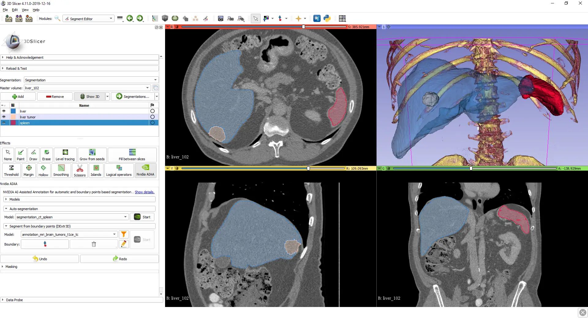
This is the oldest family and the most used in research. These are the field’s historical environments, designed for medical image analysis by a single operator. They offer complete three-dimensional visualisation, a wide set of contouring tools and extensibility through modules.Their strength is functional depth. Their limit lies in their single-user model: they natively handle neither the work queue, nor assignment, nor double reading, nor progress tracking. Those functions must be built around the tool, generally through scripts and naming conventions, which works but stays fragile.These workstations are heavily used to build the field’s reference corpora, which attests to their contouring quality. They are used in contexts where the organisation around the tool was purpose-built.Annotation platforms
The second family comes from the production world rather than the research one. Designed for production, they bring what the previous family lacks: user and rights management, task distribution, progress tracking, logging, protocol version management and sometimes integrated control mechanisms.Their limit is symmetrical. Many originate in generic image annotation and handle medical specifics poorly: volumetric navigation, geometry handling, multiple series, windowing. A platform excellent at organisation but weak at volumetric contouring yields disappointing productivity on segmentation tasks.Assistance frameworks
A third family supplies neither the interface nor the organisation but the intelligence, as a service plugging into an existing workstation.The field’s best documented framework works this way: it supports locally installed and web-based frontends, offers two active learning strategies to facilitate and speed up the training of segmentation algorithms, and provides sample interactive and non-interactive labelling applications usable directly on any given dataset. Its authors report significantly reduced annotation times on two public datasets.That service architecture is interesting because it decouples assistance from interface. It does presuppose infrastructure of its own, generally an equipped server, which puts it out of reach of a lightweight project.Contouring functions in medical image segmentation
Beyond the families, it is the available functions that determine productivity, and the field’s literature offers a stable inventory.A review lists them: semi-automatic active contour methods, boundary-points-based approaches, positive and negative click-based segmentation, as well as other semi-automatic methods such as region growing and level sets.These functions are not equally suited to every structure. Region growing and thresholding excel on homogeneous well contrasted structures, bone or aerated lung, and leak as soon as the boundary is ambiguous. Active contours suit compact structures with continuous boundaries and behave poorly on branching objects. Click-based segmentation is currently the most versatile, the annotator indicating inside and outside and correcting iteratively.One function deserves particular mention because it carries most of the gain on volumes: inter-slice interpolation. Contouring one slice in several and letting the tool produce the intermediates sharply reduces time, provided the treated slices are densified where geometry changes fast.What production requires in addition
Four functions separate a medical image segmentation tool usable in production from a contouring tool, and their absence is paid for in ancillary work.Queue management
Assigning studies, tracking progress, resuming after interruption, preventing two annotators from handling the same case. Without that function, organisation rests on a spreadsheet, which works at small scale and becomes a source of errors beyond it.Traceability
It is logged as work proceeds or not at all. Knowing who produced what, when, under which protocol version and with which control. That information is a required deliverable in regulated projects, and it is irrecoverable if it was not logged along the way.The control mechanism
Enabling double reading, arbitration, comparison of two annotations and computation of agreement metrics. A tool that does not allow two masks of the same study to be compared forces control to happen outside the system, which discourages control.Normalised export
This is the function whose absence is noticed latest and costs most. Producing masks in a constant format and geometry, with no manual intervention. It is what the publication cited in the introduction calls quantitative export, and it is what most often breaks at cohort scale.Input format and preparation
One step precedes contouring and determines part of its difficulty: preparing the data before making it available to annotators.Four operations recur. Conversion from the clinical exchange format to the working format, with the geometry preservation already noted. Series triage, a study frequently containing several acquisitions of which only one is relevant, and letting the annotator choose introduces needless variability. Default windowing, better fixed upstream than set individually. And de-identification, mandatory as soon as data leaves the site.These operations are automatable and benefit from being so, since performed manually they constitute an invisible load appearing in no costing. On a corpus of several thousand studies, preparation time can approach the annotation time of a simple structure, and a project ignoring it discovers a delay it does not attribute to its real cause.Tool selection must therefore encompass this step and not only the contouring station. An excellent medical image segmentation tool fed by artisanal preparation produces a chain whose bottleneck sits upstream of the visible work.The clinical exchange format question
One structuring choice separates two approaches to hospital integration. Some tools connect directly to image archiving systems through standard protocols, which avoids any file handling and natively preserves geometry and identifiers. Others work on exported files, which is more flexible but introduces a conversion step and a de-identification question. The first regime suits projects internal to an institution, the second suits outsourced projects, so the choice follows from the contractual configuration more than from a technical preference.Geometry, the commonest breaking point
One technical aspect of medical image segmentation deserves separate treatment, because it produces the costliest and least visible failures.The study leaves the imaging department carrying its complete geometry: pixel spacing, slice thickness and spacing, position and orientation in the patient reference frame. Most chains convert to a pivot format, and every conversion is an opportunity to lose or alter that information.Three failures recur. Loss of inter-slice spacing, which makes any later volumetric computation impossible. Axis inversion between reference frames, producing a mirrored mask, an error particularly insidious on a roughly symmetrical structure. And silent resampling, where a tool normalises the volume without returning the produced mask to the original frame.The defence does not depend on the tool chosen but on the validation protocol: annotate a structure of known asymmetric geometry, export, reimport, superimpose on the source study and verify alignment. Ten or so cases suffice, and this test must be redone at every tool version change, regressions of this kind being frequent.Automatic assistance and its loop
What models contribute has transformed productivity in medical image segmentation, and how the tool integrates it matters as much as model quality.Three integration regimes occur. Batch pre-annotation, where a model processes the whole corpus upfront and the annotator corrects, is simplest to set up and suits structures well covered by available models. Interactive assistance, where the model responds to the annotator’s indications during work, is more effective on difficult structures. And the active learning loop, where the model periodically retrains on validated annotations, is the highest performing and the most infrastructure-hungry.That third option warrants a caveat. It presupposes model and corpus evolving jointly, which complicates traceability: two studies annotated three months apart were annotated with different assistance. That must be logged, otherwise an inexplicable heterogeneity will appear in the corpus.In all three cases, anchoring remains the main limit. Faced with a plausible proposal, an annotator accepts more readily than corrects, and that tendency grows with the apparent quality of the proposal. The countermeasure lies not in the tool but in the arrangement: reserve a fraction of the corpus for unassisted work and compare the two populations.The workstation
One hardware factor is regularly underestimated although it bears directly on productivity and on quality.Navigating a volume of several hundred slices demands a responsiveness not every setup guarantees. A fraction of a second delay at each slice change, multiplied by thousands of changes, transforms a working day and pushes the annotator to work faster and worse.Three elements matter. Available memory, a three-dimensional volume occupying far more than an image. The display, the fineness of structures and the importance of windowing making screen quality non-negligible. And, in the remote configurations imposed by confidentiality, latency, which is the principal degradation factor.That last point deserves particular attention on constrained projects. A secure but slow environment produces a secure and mediocre corpus, and oversizing it costs less than correcting quality afterwards.The real cost of tooling
The licence price of a medical image segmentation tool is rarely the dominant line, and reasoning from it leads to expensive choices.Four lines make up the real cost. Setup, including integration with the preparation chain, rights configuration and geometric validation. Training, a rich tool taking several days before a team reaches its steady pace. Maintenance, versions, fixes and regressions to revalidate. And exit cost, meaning what migrating would take, which depends directly on whether the formats produced are standard or proprietary.That last line deserves evaluating before commitment rather than after. A tool producing masks in an open format and standard geometry leaves the project free; a tool whose annotations only come out in a proprietary format creates a dependency whose cost appears only when a change becomes desirable.The prudent rule is to verify, before starting, that a complete corpus can be exported to a standard format without loss and reimported elsewhere. That test costs a day and determines the project’s freedom for years afterwards.A decision grid for tooling
Six questions settle the tooling choice at project start.How many studies and how many annotators, the answer determining whether organisational functions are necessary or superfluous. Is the project regulated, which makes traceability mandatory rather than comfortable. Which structures are targeted, since the nature of useful contouring tools follows directly. Is automatic assistance available on those structures, and in what form. What confidentiality constraints bear on hosting and on the workstation. And what export format does the receiving team expect, a question to be asked before the choice rather than after.Those six answers usually point to an obvious configuration, often a combination rather than a single tool: a platform for organisation, a workstation for volumetric contouring, an assistance framework for pre-annotation.What the tool should constrain
One idea deserves reversing at the end, because it is counterintuitive and it clearly separates the projects that hold.In production, the freedom a tool offers is most often a defect. An environment letting each annotator set their windowing, choose their contouring method and name their structures their own way mechanically produces variability, whatever the quality of the written protocol.Four constraints are desirable rather than endured. Windowing fixed per task, guaranteeing everyone sees the same image. An imposed structure list with its labels, removing nomenclature divergence. Mandatory processing of cases in the assigned order, preventing cherry-picking of easy cases. And the impossibility of validating an incomplete study, avoiding silent omissions.A tool configurable to impose those four beats a richer but permissive tool in medical image segmentation production. It is a selection criterion rarely mentioned in comparisons, because it only makes sense in a multi-annotator context.What does not depend on the tool
A counterpoint is needed, since tooling is regularly invoked to explain problems that do not belong to it.No tool compensates for an absent protocol. An excellent environment in the hands of annotators without written conventions produces a corpus consistent with nobody, and that outcome is independent of the software.No tool compensates for faulty acquisition. If the structure is not visible in the study, no assistance function will make it appear.And no tool replaces the control mechanism. Double reading functions ease control but do not decide it, and a project tooled without a control strategy produces quickly and without knowing what.The practical consequence is that tool selection comes after the protocol and the quality arrangement, not before. The reverse order, common, leads to adapting the method to the software’s constraints rather than the other way round.Piloting the tooling before committing
Tool selection is one of the few medical image segmentation decisions that can be tested cheaply before it becomes expensive to reverse, and skipping that test is common.A useful pilot runs the full chain on a small number of studies rather than demonstrating the contouring interface. That means taking studies from their source format, through preparation, assignment, annotation by two different people, control, and export to the format the receiving team expects. Twenty studies are usually enough.What that pilot reveals is rarely what a demonstration shows. It surfaces the manual steps nobody mentioned, the metadata that does not survive the round trip, the fields the platform cannot record, and the real time per study once navigation and saving are included rather than contouring alone. It also produces the first measurement of inter-annotator agreement under this specific tooling, which is the only figure that predicts what production will look like.Two rules make the pilot honest. Include at least one difficult case, since tools differentiate on hard structures rather than easy ones. And have it run by the people who will do the work, not by the person evaluating the tool, whose familiarity distorts every timing.The most common mistakes
These failures recur often enough across corpus projects that naming them is usually enough to avoid them.- Choosing a tool from comparisons written for research use.
- Adopting a platform strong on organisation but weak on volumetric contouring.
- Adopting a contouring workstation without building the organisation around it.
- Not testing the conversion round trip on an asymmetric structure.
- Not redoing that test after a tool version change.
- Not logging the assistance version used in an active learning loop.
- Undersizing the remote environment imposed by confidentiality.
- Discovering the expected export format after the tool has been chosen.
- Expecting a tool to compensate for absent protocol or control.
- Choosing the tool before writing the protocol.